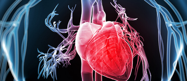
CAG Heart Surgery
Coronary angiography(CAG) of the heart
An X-ray picture of blood arteries after they have been filled with contrast material is called angiography. The “gold standard” for evaluating coronary artery disease is cardiac angiography, often known as coronary artery disease(CAD). The exact location and severity of CAD can be determined using coronary angiography.
How is it carried out?
Coronary angiography is conducted under general anesthetic with intravenous sedation and is often painless.
• A thin catheter (a thin hollow tube with a diameter of 2-3 mm) is inserted directly into an artery in the groin or arm during coronary angiography.
• The catheter is then moved to the entrance of the coronary arteries with the use of a fluoroscope (a special X-ray viewing tool) (the blood vessels supplying blood to the heart).
• A tiny quantity of radiographic contrast (an iodine-based solution that may be seen on X-ray pictures) is then injected into each coronary artery. Angiography is the name for the pictures that are created.
• It takes about 20-30 minutes to complete the treatment.
• The catheter is withdrawn after the surgery, and the artery in the leg or arm is sutured, “sealed,” or treated with manual compression to stop bleeding.
• If an angioplasty or stent is needed, it will almost always be done at the same time.
Angioplasty
A medical technique known as angioplasty is used to open up a blocked or restricted artery surrounding the heart. It is a common therapy for arteries in this part of the body that are constricted or obstructed.
The word “angioplasty” refers to the technique in which a balloon has been used to stretch a restricted or obstructed artery open. Furthermore, most advanced angioplasty procedures also include the insertion of a small wire-mesh tube into the artery, known as a stent.
To improve blood flow more freely, the stent is left in place permanently.
Angioplasty is divided into two types:
• Balloon angioplasty, which involves removing plaque from an artery using the pressure of an expanding balloon. Unless doctors are unable to insert a stent in the proper spot, this is rarely done alone.
• Stent insertion in the artery, which includes inserting a wire mesh tube, or stent, into the artery. After angioplasty, stents assist to keep an artery from narrowing again.
Stents can be formed with bare metal or with a pharmaceutical coating.
Procedure:-
A healthcare expert will clean and numb the place where the catheter enters the body, which is usually the groin but can also be the wrist, before commencing angioplasty.
The catheter is then inserted into the artery and guided towards the coronary artery while an X-ray feed monitors its progress.
After inserting the catheter, the doctor injects a contrast dye into the artery, which aids in the detection of obstructions around the heart. The doctor then inserts a second catheter and a guidewire, generally with a balloon at the tip, after locating the obstructions.
When the second catheter is in place, the doctor inflates the balloon, which pushes the plaque away from the artery and opens it up. The surgeon may place a stent in the artery to keep it open.

